Your patient today is Ling, a 43-year-old Chinese-American female who is a new patient in your practice.
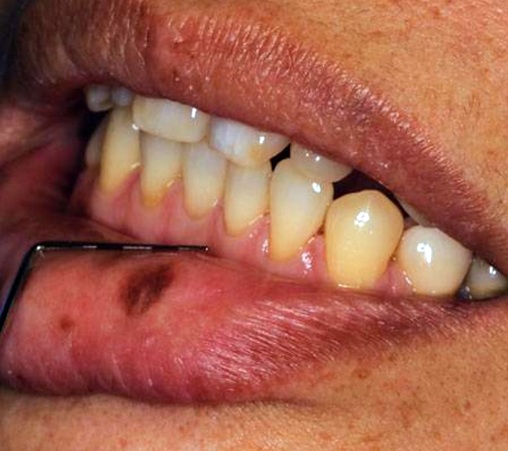
As the extraoral exam begins, you notice a pigmented macule on her lower lip.
Upon questioning her, she states that the pigmentation has been there for as long as she can remember. She reports that there has never been an issue with the pigmented area such as irritation, bleeding, or ulceration.
Ling wants to know why she has this darker pigmentation and if there is any way to remove it.
A focal pigmented lesion that occurs on the lip or the intraoral tissues is referred to as an oral melanotic macule or labial melanotic macule when occurring on the lip.
► Read also: Gravidarum granuloma associated to an osseointegrated implant: case report
The oral melanotic macule is the most common oral lesion of melanocytic origin. There is an increase due to melanin deposits in the basal cell layer, lamina propria, or both in the microscopic characteristics of the macule.
Labial melanotic macules (occurring on the lips) most often are seen in adults. But as stated in Ling’s case history, they may be present early in life.
rdhmag.com
By Nancy W. Burkhart, BSDH, EdD
Disqus comments